
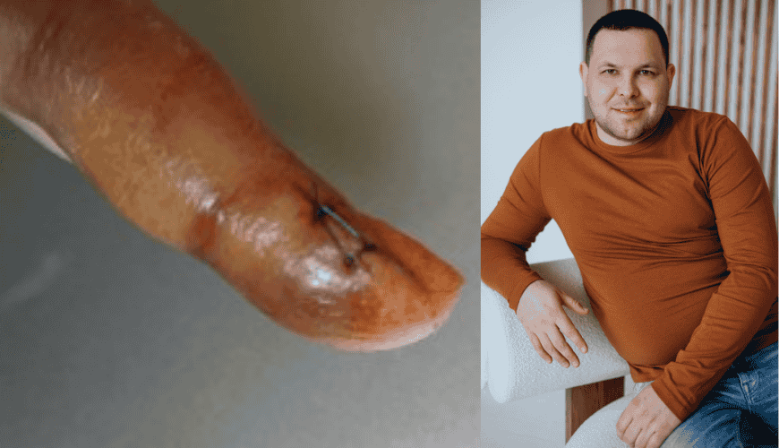
Glomus angioma is a neoplasm of mesenchymal origin, accounting for about 2% of soft tissue tumors. It is a benign tumor that originates from the neuromuscular-vascular node of the skin (glomus) due to the predominant growth of arteriovenous anastomoses.
The goal is to familiarize podiatric practitioners with glomus angiomas, the features of the course, diagnostics and treatment tactics.
Clinical cases. To illustrate the complexity of the differential diagnosis of glomus angioma, a clinical case of a 58-year-old woman is presented. It is noted that the localization of the formation is on the finger of the hand (Fig. 1.), differentiation occurred with panaritium and hygroma, the clinical and external signs of which and some morphological characteristics are very similar to vascular formations. This case demonstrates the sufficient complexity of establishing a clinical diagnosis.
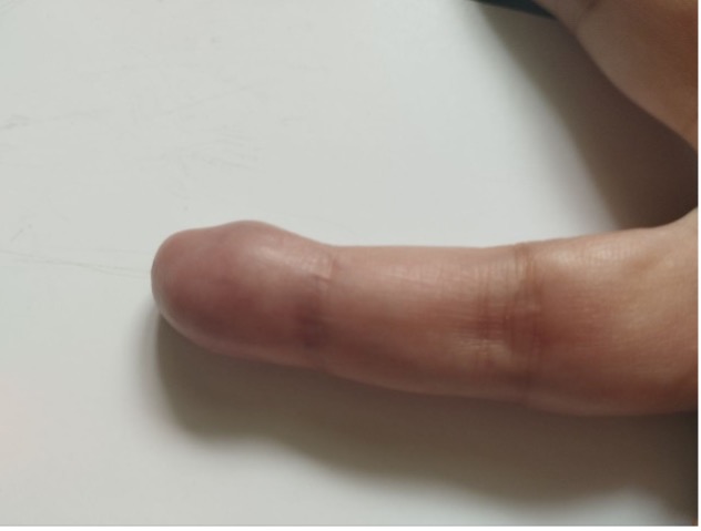
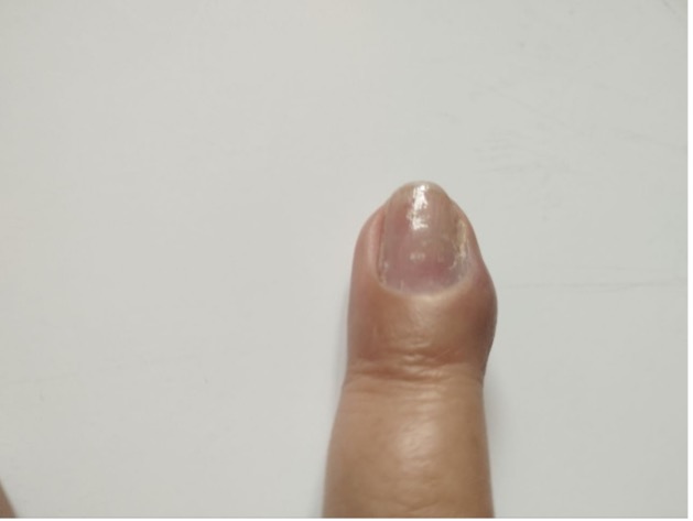
Fig. 1. General appearance of a deformed nail phalanx.
Patient M. turned to podiatrist Rita Balint with complaints of constant pain in the area of the nail phalanx of the IV finger of the left hand for 12 years, which intensifies during cold weather and is not relieved by non-narcotic painkillers. She was treated more than once by neurologists and surgeons regarding this problem. She noted periodic short-term improvement. Over time, the condition worsened, the pain intensified, and drug therapy did not help.
The patient was referred for inpatient treatment. Where she underwent the following examinations - MRI of the left cyst, coagulogram, general blood test, biochemical blood test, blood group and Rh factor.
The examination data are given below. MRI of the non-standard anatomical area of the fingers of the left cyst (Fig. 2.): on a series of MR tomograms, the bones of the fingers of the hand are not deformed. Sharpening of the edges of the articular surfaces of the finger flanks is noted. The cortical layer of the bones has a normal thickness. The articular cartilages are thin. Subchondral erosions are determined. At the level of the distal phalanx of the fourth finger, a formation is visualized that closely adheres to the bone tissue and the flexor tendon of the finger, with uneven clear contours, increased in PD, decreased in T1ZZ MR signal, measuring 0.8x0.7x1.0 cm. The tendons of the bone are without features. Soft tissues without signs of change in MR signal intensity. Along the tendons of the fingers - without fluid. Joint capsules are thickened. In the joint cavity - without fluid. Periarticular tissues without edema. CONCLUSION MR signs of arthrosis of the interphalangeal joints, tissue formation at the level of the distal phalanx of the fourth finger (probably the flexor tendon).
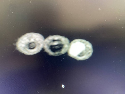
Fig. 2. MR signs of tissue formation at the level of the distal phalanx of the fourth finger (probably the flexor tendon).
Coagulogram: PTI= 87%, plasma recalcification time__ 145 sec., fibrinogen 3590 gm/l., b-naphthol test – negative, reference test – negative. Complete blood count: Hb-115 g/l., er-4.15 t/l., leuk-6.66 g/l., glucose 5.2 mmol/l. Biochemical blood test: total protein – 66.1 g\l., urea- 3.9 mmol/l., creatinine- 75.7 μmol/l., bilirubin 9.7 μmol/l. ALT-26.5 units., AST – 22.1 units. Blood group and Rh factor: O(I) first, Rh+(positive).
After establishing a preliminary diagnosis - a benign neoplasm of the IV finger of the left hand, the neoplasm was excised with subsequent histological examination (Fig. 3.)
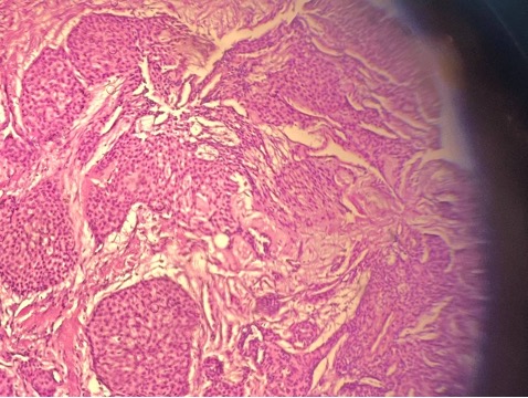
Fig. 3. Histological examination of the obtained material - glomus angioma.
The postoperative period proceeded without complications (Fig. 4.) and after removing the sutures on the 28th day (Fig. 5.) the pain completely disappeared.
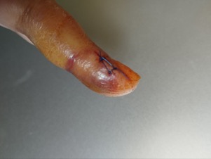
Fig. 4. Appearance on the fifth day after surgery.
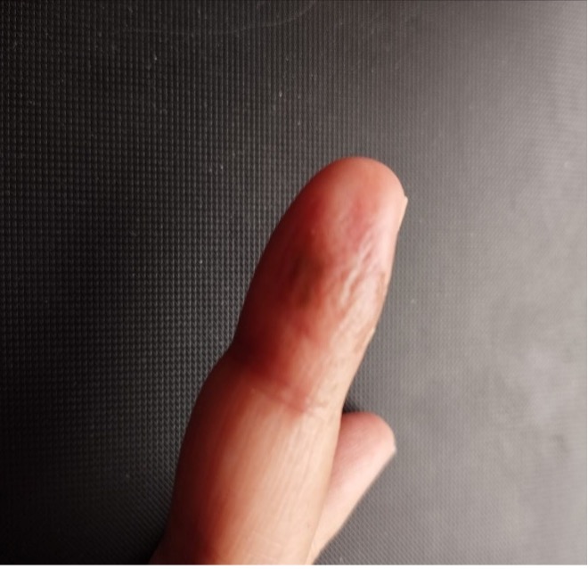
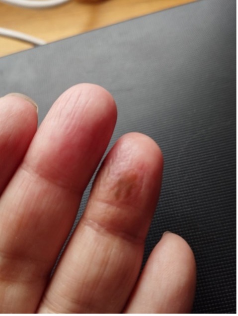
Fig. 5. Appearance 28 days after surgery.
Conclusions: According to the literature, the cause of glomus angiomas is mutations in the glomulin gene, which encodes a 68 kDa protein with an unknown function. Differential diagnosis of glomus angiomas should be carried out with pronodal hidradenoma, intradermal nevi, melanomas, myopericytomas and myofibromatosis, tumors of non-epithelial, smooth muscle, vascular or nervous origin. An important diagnostic method in the process of establishing the diagnosis is MRI and histological examination. If an adult is suspected of having a glomus angioma, with clinical symptoms and appearance appropriate for this tumor, radical removal of the formation with mandatory histological confirmation is necessary.